Pain in a joint, tendon or muscle often feels simple at first. A sore shoulder after tennis. A swollen knee after a long walk. Tingling in the hand that starts to wake you at night. Yet the real source of symptoms is not always obvious from a physical examination alone.
That is where a musculoskeletal ultrasound scan can be extremely useful. For people in Oxford, Oxfordshire and nearby areas, it offers a fast, radiation-free way to look at soft tissues, joints, nerves and fluid-filled structures in real time. It can also do something equally valuable: help guide treatment with much greater precision, especially when an ultrasound-guided injections is being considered.
Why musculoskeletal ultrasound scans matter for diagnosis
A musculoskeletal ultrasound scan, often shortened to MSK ultrasound, uses sound waves to create live images of structures just beneath the skin. Unlike an X-ray, it does not show bones particularly well, but it is excellent for tendons, ligaments, muscles, bursae, joints, nerves and cysts. That makes it especially relevant for many of the painful conditions seen every day in specialist MSK clinics.
This type of scan is often used when someone has pain, swelling, weakness, clicking, a lump, pins and needles or loss of movement. NHS guidance also recognises ultrasound as a way to investigate pain and lumps, and to help clinicians carry out procedures including steroid injections into joints. In practice, that means it can support both diagnosis and treatment planning in a single pathway.
A major strength of MSK ultrasound is its immediacy. The clinician can scan the exact area that hurts, press on it, ask you to move, and compare the painful side with the unaffected side. That often helps connect the image to the symptom rather than simply producing a static picture.
After an initial assessment, a scan is often valued because it can:
- show soft tissue injury in real time
- identify fluid, inflammation or swelling
- assess movement while a joint or tendon is working
- help decide whether rehabilitation, injection treatment or other care is the right next step
What an MSK diagnostic ultrasound scan can identify
The phrase “musculoskeletal ultrasound” covers a wide range of findings. It can spot tendon damage, inflammation around tendons, bursitis, joint fluid, synovial thickening, nerve compression, ganglion cysts and some soft tissue lumps. In the hands and wrists, high-frequency ultrasound has long been recognised as a quick and efficient way to assess soft tissue disease, including tendon injuries, annular pulley problems, ligament damage and tenosynovitis.
The table below shows the sort of conditions that may be identified during an MSK diagnostic scan.
| Body area | Common ultrasound findings | Why it matters |
|---|---|---|
| Shoulder | Rotator cuff tears, bursitis, calcific tendinopathy, joint effusion | Helps separate tendon pain from joint or bursal pain |
| Elbow | Tennis elbow changes, golfer’s elbow changes, tendon tears, bursitis, nerve irritation | Useful when pain persists despite rest or exercise |
| Wrist and hand | Tenosynovitis, ganglion cyst, carpal tunnel features, tendon injury, localised swelling | Often very effective for small soft tissue structures |
| Hip and groin | Bursitis, tendon pathology, joint effusion, snapping structures | Helps narrow down anterior hip or lateral hip pain |
| Knee | Effusion, Baker’s cyst, tendon problems, bursitis, signs linked with osteoarthritis | Useful when swelling or anterior knee pain is present |
| Ankle and foot | Achilles tendinopathy, plantar fasciitis, ligament injury, Morton’s neuroma, tendon tears | Valuable for runners and people with persistent foot pain |
In inflammatory or irritated joints, ultrasound can identify changes including synovial hypertrophy, which means thickening of the joint lining, and joint effusion, which means excess fluid. Those findings matter because they can point to active inflammation and can help explain stiffness, swelling and pain.
In specialist MSK practice across Oxfordshire, this is especially helpful for common problems involving the shoulder, knee, ankle and hand, where a careful scan can often clarify whether the pain is coming from a tendon, a joint, a bursa or an irritated nerve.
Soft tissue, joint, nerve and cyst problems seen on ultrasound
Tendon pathology is one of the most common reasons to request a scan. Ultrasound can show tendinopathy, partial tears, full-thickness tears and inflammation around the tendon sheath, known as tenosynovitis. In the shoulder, that may involve the rotator cuff. In the ankle, it may involve the Achilles, peroneal or tibialis posterior tendons. In the hand, it may reveal stenosing tenosynovitis or trauma-related injury.
Joint-related findings are another major area. A scan can detect joint effusion, inflamed synovium, bursitis and some arthritic changes. While it does not replace every other type of imaging, it is very useful when the question is whether there is active inflammation, fluid or a structure that is being irritated during movement.
Nerve assessment is also highly relevant. Ultrasound can help assess nerve entrapment syndromes, including carpal tunnel syndrome at the wrist and ulnar nerve irritation around the elbow. It can also help identify whether a nearby cyst, swelling or soft tissue change is contributing to compression.
Localised lumps can often be assessed very effectively with ultrasound. Ganglion cysts are a good example, especially around the wrist or foot. The scan can also help distinguish a fluid-filled structure from a more solid soft tissue lump, which is a very practical step when deciding what should happen next.
When clinicians talk about what a musculoskeletal ultrasound can pick up, they are often referring to categories like these:
- Tendon problems: tendinopathy, partial tears, full tears, tenosynovitis
- Joint changes: synovial hypertrophy, joint effusion, bursitis, osteoarthritic irritation
- Nerve issues: nerve entrapment, local nerve swelling, compression from nearby tissue
- Cyst and lump assessment: ganglion cysts, fluid collections, soft tissue swelling
Why real-time ultrasound imaging helps in Oxfordshire sports and arthritis care
One of the biggest practical advantages of ultrasound is that it is dynamic. The scan is performed while the patient can move the painful area, which means the clinician may see structures catching, snapping, subluxing or becoming painful under load. That is hard to reproduce with static imaging alone.
This is particularly relevant in active communities across Oxford, Abingdon, Witney, Bicester, Didcot and the wider Oxfordshire area. Runners often present with Achilles pain or plantar fascia symptoms. Cyclists may develop hip or knee overload patterns. Racquet sport players may struggle with shoulder or elbow pain. A live scan can add detail that supports a more accurate rehabilitation plan.
The same applies to arthritis care. If a knee is swollen, a scan can show whether the swelling is truly inside the joint, whether there is associated synovitis, and whether there are neighbouring structures involved, including a Baker’s cyst. That makes the scan not just diagnostic, but clinically useful in deciding whether physiotherapy, aspiration, guided injection or another approach makes sense.
Ultrasound-guided injections after MSK diagnostic scans
A good MSK pathway does not stop at naming the problem. It should help direct the next step with confidence. This is where ultrasound-guided injections are especially important.
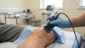
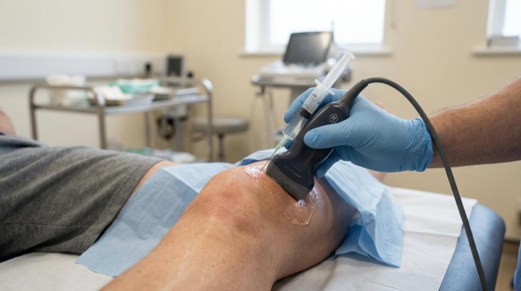
Using ultrasound guidance, the clinician can see the needle in real time and place the treatment into the intended structure with much greater accuracy. NHS guidance recognises this use clearly, including helping inject a steroid into a joint. That matters because the target may be small, deep or close to important nearby structures.
In practical terms, a diagnostic scan often answers several key questions before an injection is offered. Is the pain source actually inflamed? Is there fluid in the joint? Is the bursa the main problem, or is the tendon the real driver? Is the person’s pain pattern consistent with the structure being treated? Those details improve decision-making and help keep treatment focused.
The most commonly discussed ultrasound-guided injection options include:
- Steroid injections: often used when inflammation is a major part of the problem, including bursitis, synovitis, some arthritic flares and tenosynovitis
- Hyaluronic acid injections: often considered for osteoarthritic joints, especially when lubrication and cushioning within the joint are part of the treatment goal
- Arthrosamid injections: a non-surgical injectable option sometimes considered for knee osteoarthritis after careful clinical and imaging assessment
Steroid injections are often most helpful when there is a clear inflammatory target. A scan can show joint effusion, synovial thickening or bursal swelling, making it easier to decide whether a steroid injection is likely to be relevant. It can also help guide injections around the shoulder, wrist, hip, knee, ankle and foot with greater precision.
Hyaluronic acid injections are typically discussed in the setting of joint degeneration, especially knee osteoarthritis. If the scan supports the view that the joint itself is a meaningful pain source, accurate placement into the joint becomes a priority. Ultrasound guidance supports that precision.
Arthrosamid injections have become a talking point for people seeking a non-surgical approach to persistent knee osteoarthritis. Not every knee pain case is suitable, and not every arthritic knee is painful for the same reason. A proper MSK assessment and diagnostic imaging help confirm whether the joint is the key pain generator before this type of treatment is considered. For patients in Oxford and surrounding areas weighing up different knee injection options, that clarity is essential.
GB Clinics provides MSK diagnostic ultrasound scans alongside ultrasound-guided injection therapies across shoulder, elbow, wrist and hand, hip, knee, ankle and foot conditions. In specialist settings, this joined-up approach can reduce delay between assessment, diagnosis and targeted treatment.
When to book a musculoskeletal ultrasound scan in Oxford or Oxfordshire
People often wait longer than necessary because they assume pain just needs more time. Sometimes that is true. Sometimes it is not. A scan becomes more useful when symptoms are lingering, recurring or not responding as expected to rest, exercise or manual treatment.
It can also be appropriate when the diagnosis remains uncertain after a clinical examination. A painful shoulder may be rotator cuff related, but it could also involve bursitis, joint irritation or a combination of problems. That overlap is one reason shoulder management can be nuanced, and Rygklinikken’s evidensbaseret guide to skuldersmerter reflects how treatment choices often depend on identifying the main pain-generating structure first. Numbness in the hand may suggest carpal tunnel syndrome, yet a scan may also reveal tendon sheath inflammation or a nearby ganglion cyst.
Across Oxfordshire, access to specialist MSK assessment can be especially helpful for people who need to stay active for work, sport or daily independence. That includes office workers with repetitive hand pain, parents carrying young children, older adults trying to keep mobile, and athletes wanting clear answers before returning to training.
A scan is often worth considering if you have:
- persistent tendon or joint pain
- visible swelling or a lump
- repeated flare-ups of arthritis-type symptoms
- numbness, tingling or suspected nerve entrapment
- pain that may need an ultrasound-guided injection
What happens during an MSK ultrasound scan and what comes next
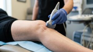
The scan itself is usually straightforward. Gel is placed on the skin, and a small handheld probe is moved over the painful region. The appointment may include movement testing during the scan, pressure over the area of symptoms and comparison with the opposite side. Because the images are live, the clinician can often explain findings as the scan takes place.
That immediate feedback is one of the strongest parts of the process. Rather than waiting days for a report before the treatment plan starts to make sense, patients can often leave with a clearer picture of what has been found and what should happen next. That next step may be physiotherapy, load modification, a rehabilitation programme, shockwave therapy, podiatry input, or an ultrasound-guided injection.
For people in Oxford, Oxfordshire and nearby towns, the real value of MSK diagnostic ultrasound is not just that it can identify a problem. It is that it can connect symptoms, imaging and treatment in a practical way, whether the issue is a rotator cuff tear, tenosynovitis, synovial irritation, a ganglion cyst, plantar fasciitis, carpal tunnel features or an osteoarthritic knee being considered for steroid, hyaluronic acid or Arthrosamid injection treatment.