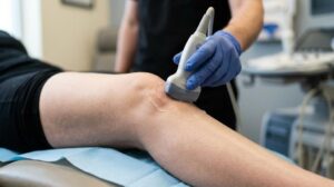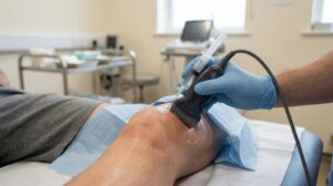

What if the reason your persistent joint pain remains a mystery isn’t because there’s no solution, but because your previous scans only captured a still image of a moving problem? It’s common to feel frustrated by vague diagnoses like “wear and tear” or to feel anxious about the confined space of an MRI tunnel. You deserve a definitive answer that doesn’t rely on invasive procedures or radiation. This is where a diagnostic ultrasound musculoskeletal assessment changes the narrative, offering a dynamic view of your body in motion.
In this complete 2026 guide, you’ll discover how this advanced technology provides real-time, radiation-free insights into your pain to accelerate your recovery. We’ll explain how these scans lead to immediate answers during your appointment and facilitate a personalised, non-surgical treatment plan. From guided steroid injections to shockwave therapy, you’ll learn how we use precise imaging to move you away from uncertainty and towards active, pain-free living.
Key Takeaways
- Understand how high-frequency sound waves provide a safe, radiation-free window into your soft tissues for a clearer, more precise diagnosis.
- Learn why a diagnostic ultrasound musculoskeletal assessment often reveals finer detail in small tendons and ligaments than traditional imaging methods.
- Discover the benefits of dynamic scanning, where moving the joint during the procedure helps trigger and identify the exact source of your symptoms in real-time.
- Identify common conditions such as tendon tears, bursitis, and joint inflammation that can be diagnosed immediately during your clinical consultation.
- See how accurate imaging forms the foundation for targeted, non-surgical treatments like ultrasound-guided injections to ensure medication reaches the precise area of injury.
Understanding Diagnostic Musculoskeletal (MSK) Ultrasound
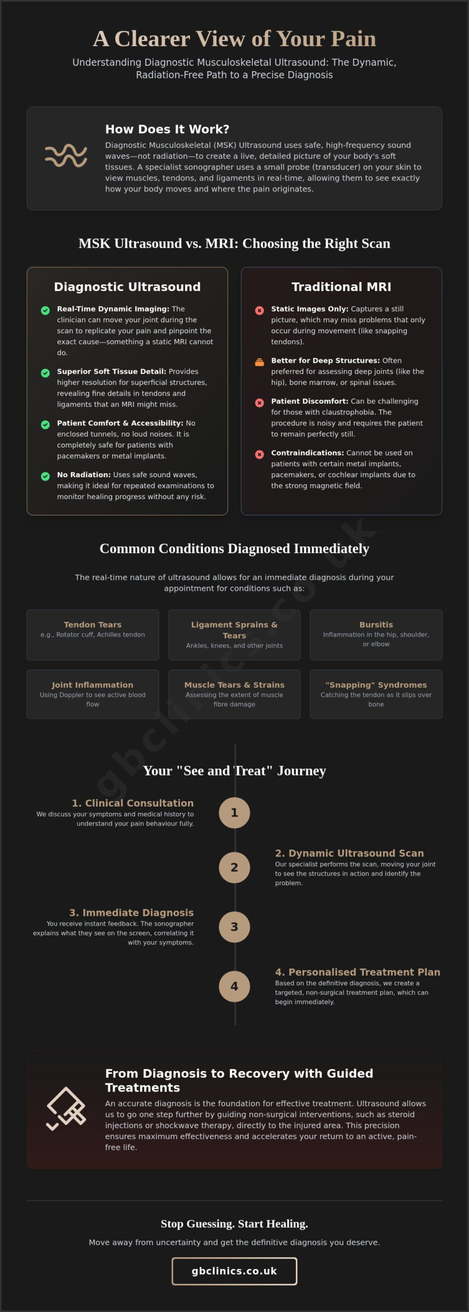
Diagnostic ultrasound musculoskeletal imaging serves as a sophisticated tool for assessing the body’s intricate soft tissue structures. Unlike traditional radiography that focuses primarily on bone density, this technology uses high-frequency sound waves to generate high-resolution images of tendons, ligaments, and muscles. These sound waves bounce off internal structures and return to a transducer, creating a visual map of the area of concern. This process, known as Medical ultrasonography, allows clinicians to observe the internal landscape of a joint without the need for invasive measures or ionising radiation.
The most significant advantage of this modality is its dynamic nature. Most medical scans require you to remain perfectly still, but an MSK ultrasound thrives on movement. We can observe how a tendon glides or how a muscle contracts in real-time. This provides a functional assessment that static images simply cannot match. It’s often the difference between seeing a “normal” joint and discovering the exact moment a ligament catches or a tendon pinches during a specific movement.
The Role of the Specialist Sonographer
A successful scan depends heavily on the expertise of the clinician holding the probe. Musculoskeletal imaging requires a higher level of specialised training than general abdominal scans because the anatomy is incredibly complex. Our specialists in Oxfordshire don’t just look at the screen; they perform a clinical correlation. This means they match what they see on the monitor with your specific pain behaviour. By discussing your history during the diagnostic ultrasound musculoskeletal assessment, they provide immediate feedback, ensuring the diagnosis aligns with your physical experience.
Safety and Accessibility of Ultrasound Imaging
Safety is a primary concern for many patients, especially those who have undergone multiple diagnostic tests. Because ultrasound uses sound waves rather than radiation, it’s entirely safe for repeat examinations. This makes it an ideal choice for monitoring the progress of a recovery plan or checking the healing status of a muscle tear. There’s no cumulative risk, which provides peace of mind for patients requiring long-term care.
Accessibility is another key benefit. Patients with cardiac pacemakers, cochlear implants, or metal fragments in their bodies often cannot undergo MRI scans due to the powerful magnetic fields involved. Ultrasound presents no such risks. In our private clinic centre, we embrace a “see and treat” philosophy. This streamlined approach means you can move from a diagnostic scan to a targeted treatment plan, such as a guided steroid injection, in a single, efficient journey.
MSK Ultrasound vs MRI: Why Real-Time Imaging Wins
Choosing between an MRI and a diagnostic ultrasound musculoskeletal scan often depends on the specific nature of your injury. Whilst many patients assume an MRI is the only way to get a clear picture, ultrasound actually provides superior resolution for superficial structures. It can capture the fine, individual fibres of a tendon that a standard MRI might miss. This high level of detail is vital when we’re looking for tiny tears or subtle changes in tissue health. It’s not just about seeing the anatomy; it’s about seeing it with incredible clarity.
Patient comfort is another area where ultrasound excels. You don’t have to worry about the claustrophobia or the loud, rhythmic clanging associated with MRI tunnels. Instead, you’ll be in a comfortable, open environment where you can speak directly with your clinician. In the UK, private wait times for an MRI can often stretch into weeks, but a diagnostic ultrasound musculoskeletal assessment is typically much more accessible. This speed allows us to move you from pain to a recovery plan without unnecessary delays. If you’re ready to find answers, a clinical consultation is your first step towards a precise diagnosis.
When Ultrasound is the Superior Choice
Ultrasound is the gold standard for diagnosing “snapping” or “popping” sensations. Because we can move the joint whilst scanning, we can catch the exact moment a tendon catches on a bone or a ligament slips. We also use Doppler technology to assess blood flow, which is a powerful way to detect active inflammation. This patient guide to musculoskeletal ultrasound highlights how effective the technology is for assessing the Achilles tendon, the rotator cuff, and other structures close to the skin’s surface.
The Limitations: When an MRI Might Still Be Needed
There are instances where we’ll recommend an MRI instead. Ultrasound waves cannot penetrate bone, so we can’t use it to see deep inside a joint to check the ACL or the deep labrum of the hip. It also isn’t suitable for assessing bone marrow oedema or internal bone health. If our initial assessment suggests your issue is buried deep within the joint or involves the bone itself, we’ll always advise you honestly on which imaging modality will provide the most reliable answers for your specific case.
Common Conditions Diagnosed Through MSK Ultrasound
A diagnostic ultrasound musculoskeletal assessment provides a detailed look at the structures that keep you moving. It is particularly effective at identifying pathologies that might be missed by other imaging methods or physical examinations alone. By visualising the intricate relationship between soft tissues, we can accurately diagnose conditions ranging from acute sports injuries to chronic degenerative changes. This precision is essential because the right treatment depends entirely on an accurate understanding of the underlying pathology.
Clinicians frequently rely on this modality to investigate tendon health. Whether it’s identifying a small tear, chronic tendinopathy, or the presence of calcific deposits, ultrasound offers a clear view of the tissue’s internal architecture. It’s also an invaluable tool for detecting joint inflammation, such as synovitis or bursitis in the shoulder, hip, and knee. According to an NIH review on musculoskeletal ultrasound, the ability to assess these conditions in real-time makes it a cornerstone of modern orthopaedic diagnostics.
Beyond tendons and joints, we use ultrasound to evaluate:
- Ligament Sprains: Assessing the severity of injuries in the ankle or wrist to determine if a tear is partial or complete.
- Nerve Entrapment: Visualising nerves to diagnose conditions like Carpal Tunnel Syndrome or Morton’s Neuroma, where we can see physical compression or swelling of the nerve.
- Muscle Tears: Localising the exact site and extent of a muscle strain to provide an accurate recovery timeline.
Shoulder and Rotator Cuff Assessments
The shoulder is perhaps where ultrasound shines brightest. It’s widely considered the gold standard for diagnosing rotator cuff tears, as it allows us to test the integrity of the tendons throughout their full range of motion. We can easily identify subacromial bursitis, a common cause of impingement pain that often feels like a sharp catch when you lift your arm. We also frequently diagnose calcific tendonitis, which is the accumulation of calcium hydroxyapatite crystals within the tendon fibres that ultrasound identifies as distinct, bright deposits.
Knee and Ankle Soft Tissue Injuries
In the lower limbs, ultrasound is equally effective. We can quickly check for Baker’s cysts, which are fluid-filled sacs that develop behind the knee and cause significant discomfort. For ankle issues, we focus on the Achilles tendon, distinguishing between inflammation and partial or full-thickness tears. We also measure the thickness of the plantar fascia; this data is crucial for tailoring our shockwave therapy or guided injection protocols to your specific needs.
What to Expect During Your Appointment at GB Clinics
Your journey at GB Clinics begins with a comprehensive clinical consultation rather than just a technical scan. We believe that a diagnostic ultrasound musculoskeletal assessment is most effective when it’s informed by your specific story. Our specialists spend the first part of your appointment discussing your pain behaviour, previous injuries, and how your symptoms affect your daily life. This dialogue ensures that when the probe touches your skin, we’re looking for answers in the right places. It’s a methodical approach that combines clinical expertise with advanced technology to provide a complete picture of your health.
The scanning process itself is straightforward and comfortable. We apply a cool, water-based gel to the area of concern, which helps the sound waves travel from the transducer probe into your soft tissues. As the clinician moves the probe across your skin, high-resolution images appear instantly on a monitor. One of the most reassuring aspects of our service is the open communication. Your clinician will explain exactly what you’re seeing on the screen as it happens. You won’t be left wondering about the results; you’ll receive a clear explanation of the findings before you even leave the room.
Efficiency is a cornerstone of our care. In many traditional medical settings, you might wait days or even weeks for a radiologist to send a report back to your doctor. At our clinic, the results are immediate. This allows us to move straight from diagnosis to discussing your recovery options. If you’re ready to stop guessing about your joint pain, you can book your diagnostic assessment today and get the answers you need in a single visit.
Preparing for Your Scan
Preparation for your appointment is minimal. We recommend wearing loose, comfortable clothing that allows easy access to the joint being scanned. If we’re assessing a shoulder, a vest top is often helpful, whilst shorts are ideal for knee or ankle examinations. Most diagnostic sessions take between 20 to 30 minutes, providing ample time for a thorough investigation without feeling rushed. You don’t need a GP referral to access our private services in Oxfordshire; you can simply reach out to us directly to organise your visit.
The Interactive Nature of the Scan
What sets this experience apart is how interactive it feels. Your clinician might ask you to perform a specific movement or contract a muscle to “reproduce the pain” whilst they watch the tissues on the screen. This dynamic interaction is often the key to finding the source of a problem that only occurs during activity. Seeing your own anatomy in motion can be incredibly empowering. It replaces the anxiety of the unknown with a tangible understanding of your body, making the path to recovery feel much more achievable.
From Diagnosis to Recovery: Ultrasound-Guided Treatments
A precise diagnosis is the bedrock of any successful recovery. It’s the difference between treating a general area of discomfort and addressing the specific root cause of your pain. A diagnostic ultrasound musculoskeletal assessment provides the clarity required to move forward with confidence. We don’t just find the source of your pain; we use that visual map to guide our interventions, ensuring every treatment is delivered with clinical precision. This transition from imaging to action is what allows for a faster, more effective return to health.
By using the same high-resolution technology from your initial scan, we can perform ultrasound-guided injections that target the exact site of injury. Whether we are delivering guided steroid injections for acute inflammation or prolotherapy to stimulate tissue repair, the goal is always the same: maximum accuracy with minimal discomfort. This approach also allows us to integrate other non-surgical options, such as shockwave therapy or advanced physiotherapy, based on the specific structural findings of your scan.
The Advantage of Guided Steroid and Gel Injections
Traditional “blind” injections rely on anatomical landmarks and a clinician’s intuition. Whilst experienced practitioners can be accurate, complex joints like the hip or small spaces in the wrist present significant challenges. Research suggests that blind injections have higher failure rates in these areas compared to those performed under guidance. By visualising the needle tip in real-time, we can navigate around nerves and blood vessels, ensuring the medication reaches the target tissue. This is particularly effective for our Guided Hyaluronic Acid Injection service, where the gel must be placed precisely within the joint space to provide optimal lubrication and pain relief.
A Holistic Approach to Musculoskeletal Health
Our commitment to your recovery extends far beyond the initial scan. We use diagnostic insights to help our clinicians organise your rehabilitation journey. If a follow-up scan shows a muscle tear is healing well, we can safely progress your loading exercises in physiotherapy. Conversely, if the tissue requires more stimulation, we might introduce shockwave therapy to encourage new blood vessel growth. This Oxfordshire-based care ensures you have professional support from your first diagnostic ultrasound musculoskeletal scan through to your final stage of recovery. We focus on a steady, methodical path that prioritises your long-term physical well-being and prevents the recurrence of injury.
Take Control of Your Recovery Journey
Understanding the root cause of your pain is the most vital step towards reclaiming your mobility. By opting for a diagnostic ultrasound musculoskeletal assessment, you move beyond the uncertainty of vague diagnoses and into a clear, actionable recovery plan. This technology doesn’t just show us your anatomy; it lets us witness your body in motion, ensuring that even the most subtle injuries are identified with precision. Whether you’re dealing with a complex rotator cuff tear or chronic joint inflammation, the combination of real-time imaging and specialist expertise provides a level of detail that static scans simply cannot match.
At GB Clinics, we combine state-of-the-art ultrasound technology with specialist-led assessments to deliver immediate results and personalised treatment planning. You don’t have to wait weeks for answers or settle for “blind” treatments that may not hit the mark. Our team is dedicated to providing the clarity and support you need to return to the activities you love. If you’re ready to find a definitive solution for your persistent pain, Book Your Diagnostic Ultrasound Appointment at GB Clinics Oxford. We’re here to guide you from your first scan through to a full, active recovery.
Frequently Asked Questions
Is a musculoskeletal ultrasound as accurate as an MRI?
Accuracy depends on the specific structure being examined. For superficial tissues like the rotator cuff or Achilles tendon, ultrasound often provides higher resolution than a standard MRI. It also offers the benefit of dynamic scanning to see tissues in motion. However, for deep internal joint structures like the ACL or bone marrow health, an MRI remains the preferred diagnostic tool for most clinicians.
Do I need a referral from my GP for a private diagnostic ultrasound?
You don’t need a GP referral to access our specialist services in Oxfordshire. We accept self-referrals, which allows you to bypass long waiting lists and gain immediate insights into your pain. This direct access route is designed to streamline your journey from initial discomfort to a structured recovery plan without unnecessary administrative delays or prolonged periods of uncertainty.
How much does a private diagnostic ultrasound cost in the UK?
The cost of a diagnostic ultrasound musculoskeletal scan varies depending on the complexity of the joint and whether multiple areas require assessment. Whilst industry prices across the UK fluctuate based on the clinic’s location and the clinician’s expertise, we maintain a transparent approach to our fees. We recommend contacting our clinic centre directly to receive a specific quote tailored to your diagnostic requirements.
Can ultrasound see through bone to find fractures?
Ultrasound waves cannot penetrate the hard surface of the bone. Whilst it’s excellent for identifying issues on the bone’s surface, such as cortical irregularities or stress fractures, it cannot visualise the internal bone structure. If a deep fracture or bone marrow issue is suspected, an X-ray or MRI is usually the most appropriate imaging modality to ensure a comprehensive diagnosis.
Will the clinician explain the results to me during the scan?
Yes, our specialists provide real-time feedback throughout the entire procedure. You’ll be able to see the high-resolution images on the screen whilst the clinician explains the findings and how they correlate with your specific symptoms. This interactive approach ensures you leave your appointment with a clear understanding of your condition and the potential next steps for your treatment and recovery.
Are there any side effects or risks associated with MSK ultrasound?
There are no known side effects or risks associated with this type of medical imaging. Unlike X-rays or CT scans, ultrasound uses high-frequency sound waves rather than ionising radiation to create images. It’s a non-invasive and safe procedure for everyone, including patients with cardiac pacemakers, metal implants, or those who may be pregnant and wish to avoid radiation exposure.
How long will I have to wait for an appointment at the Oxford clinic?
We strive to offer appointments promptly, often within a few days of your initial enquiry. This responsiveness is a core part of our commitment to active recovery and patient support. You can check our current availability by contacting the clinic directly. We prioritise efficient access to ensure you don’t have to manage persistent pain without a clear diagnosis for longer than necessary.
Can I have a guided injection on the same day as my diagnostic scan?
It’s often possible to provide a guided injection on the same day as your diagnostic scan as part of our “see and treat” approach. This depends entirely on the clinical findings and whether the clinician determines that an injection is the most appropriate next step for your condition. Combining these sessions can accelerate your relief and reduce the number of visits required to the clinic.